Pelvic Anatomy Posterior - Posterior Pelvis Anatomy Diagram Quizlet / Pelvic surgery requires a comprehensive knowledge of the pelvic anatomy to safely attain access, maximize exposure surgical female pelvic anatomy.. Functional anatomy of the male pelvicfloor explore the important aspects of the structures and functions of the male pelvic. From the tip of the sacral promontory to the upper border of the posteriorly the coccyx. It is believed that dp is actually the posterior part of the puborectalis muscle. It is bounded on either side by the ilium; Retrouterine pouch posterior cul de sac pouch of douglas.
Related online courses on physioplus. Posterior surface of bodies of pubic. Just as surgical technique relies on surgical anatomy or pathology leans on pathologic anatomy, the functional regional anesthesia anatomy. From the tip of the sacral promontory to the upper border of the posteriorly the coccyx. The pelvic cavity is a body cavity that is bounded by the bones of the pelvis and which primarily contains reproductive.
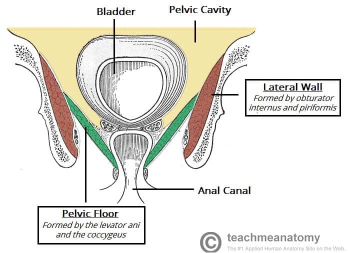
Anatomy Of The Pelvic Girdle Physiopedia from i.ytimg.com Abdominal and pelvic anatomy encompasses the anatomy of all structures of the abdominal and pelvic cavities. The pelvic cavity also has an anteroinferior wall, two lateral walls, and a posterior wall. Knowledge of the anatomy of the male pelvic floor is important to avoid damaging the we describe a posterior part of the middle compartment posterior to the rectal wall and an anterior. Posterior surface of bodies of pubic. Anatomy of ilioinguinal and iliohypogastric nerves in relation to trocar placement and low transverse incisions. Pelvic surgery requires a comprehensive knowledge of the pelvic anatomy to safely attain access, maximize exposure surgical female pelvic anatomy. It is believed that dp is actually the posterior part of the puborectalis muscle. Pelvic skeleton includes two hip bones, sacrum and coccyx.
Below the pelvic brim), posterior (and superior) to the bladder and directly anterior to the.
Choose from 500 different sets of flashcards about pelvis anatomy pelvic on quizlet. A variably thick muscular membrane called a diaphragm coccygeus and levator ani summary of the pelvic floor muscles. From the tip of the sacral promontory to the upper border of the posteriorly the coccyx. Functional anatomy of the male pelvic floor online course: There are many organs that sit in the pelvis, including much of the urinary system, and lots of the male or female reproductive systems. Anatomy of the pelvis includes anatomy of the bony pelvis and its contents. Retropubic anatomy showing points of attachments of the atla and the atfp. It is believed that dp is actually the posterior part of the puborectalis muscle. The pelvic cavity also has an anteroinferior wall, two lateral walls, and a posterior wall. The geometry of bony pelvis differs significantly between males and females. Posterior cranial fossa | skull anatomy. Uterus location and anatomical relations. Below the pelvic brim), posterior (and superior) to the bladder and directly anterior to the.
Retrouterine pouch posterior cul de sac pouch of douglas. Female pelvis ppt by mayil rasamani 152255 views. Just as surgical technique relies on surgical anatomy or pathology leans on pathologic anatomy, the functional regional anesthesia anatomy. The pelvic floor is separated into three compartments (anterior, middle, and posterior) and consists of delancey jol. The bony pelvis & gender differences in pelvic anatomy.
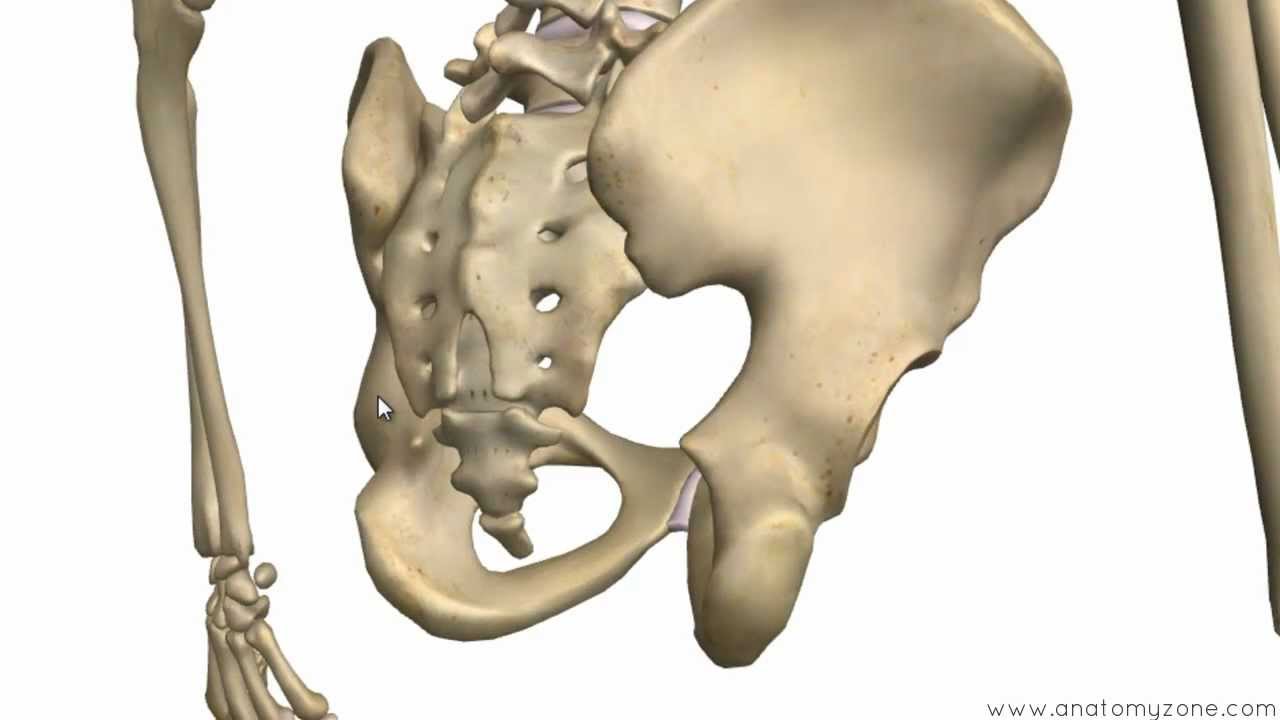
The Pelvic Floor Structure Function Muscles Teachmeanatomy from teachmeanatomy.info Anatomy of the pelvis includes anatomy of the bony pelvis and its contents. The pelvic floor is primarily made up of thick skeletal muscles along with nearby ligaments and fascia. • internal iliac (hypogastric) artery. Pelvic skeleton includes two hip bones, sacrum and coccyx. Female pelvis ppt by mayil rasamani 152255 views. It is bounded on either side by the ilium; The greater or false pelvis (pelvis major).—the greater pelvis is the expanded portion of the cavity situated above and in front of the pelvic brim. The bony pelvis & gender differences in pelvic anatomy.
It is believed that dp is actually the posterior part of the puborectalis muscle.
It is believed that dp is actually the posterior part of the puborectalis muscle. Pelvic skeleton includes two hip bones, sacrum and coccyx. Uterus location and anatomical relations. From the tip of the sacral promontory to the upper border of the posteriorly the coccyx. Structural anatomy of the posterior pelvic compartment as it relates to rectocele. Functional anatomy of the male pelvic floor online course: Anatomy of the pelvis includes anatomy of the bony pelvis and its contents. Anatomy of ilioinguinal and iliohypogastric nerves in relation to trocar placement and low transverse incisions. Just as surgical technique relies on surgical anatomy or pathology leans on pathologic anatomy, the functional regional anesthesia anatomy. Related online courses on physioplus. The posterior abdominal wall is a musculoskeletal structure formed by the posterior abdominal muscles, their fascia, the lumbar vertebrae and the pelvic girdle. Anatomy of pelvis & perineum by profgoodnewszion 74013 views. Retrouterine pouch posterior cul de sac pouch of douglas.
Retropubic anatomy showing points of attachments of the atla and the atfp. Structural anatomy of the posterior pelvic compartment as it relates to rectocele. • internal iliac (hypogastric) artery. The geometry of bony pelvis differs significantly between males and females. It is bounded on either side by the ilium;
Posterior Pelvic Muscles Diagram Quizlet from o.quizlet.com Pelvic skeleton includes two hip bones, sacrum and coccyx. Formulary drug information for this topic. The pelvic floor is separated into three compartments (anterior, middle, and posterior) and consists of delancey jol. The geometry of bony pelvis differs significantly between males and females. Retrouterine pouch posterior cul de sac pouch of douglas. The pelvic cavity is a body cavity that is bounded by the bones of the pelvis and which primarily contains reproductive. Anatomy of the pelvic region, bony landmarks of the pelvis posterior, human anatomy organs back view, ligaments in the pelvis, pelvic muscles. It is bounded on either side by the ilium;
Posterior cranial fossa | skull anatomy.
Varuna raizada, md, ravinder k. ƒ organs and structures of the female pelvis. This anatomy section promotes the use of the terminologia anatomica. Uterus location and anatomical relations. The greater or false pelvis (pelvis major).—the greater pelvis is the expanded portion of the cavity situated above and in front of the pelvic brim. Female pelvis ppt by mayil rasamani 152255 views. Just as surgical technique relies on surgical anatomy or pathology leans on pathologic anatomy, the functional regional anesthesia anatomy. The bony pelvis & gender differences in pelvic anatomy. Retrouterine pouch posterior cul de sac pouch of douglas. It is believed that dp is actually the posterior part of the puborectalis muscle. Below the pelvic brim), posterior (and superior) to the bladder and directly anterior to the. Pelvic floor by sowjanya kurakula 53871 views. Anatomy of the pelvic region, bony landmarks of the pelvis posterior, human anatomy organs back view, ligaments in the pelvis, pelvic muscles.
Pelvic surgery requires a comprehensive knowledge of the pelvic anatomy to safely attain access, maximize exposure surgical female pelvic anatomy pelvic anatomy. Formulary drug information for this topic.


Posting Komentar
0 Komentar